
Ion exchange chromatography (IEX, also written IEC) separates molecules by charge. Analytes bind to a stationary phase carrying the opposite charge, then release when you raise the salt concentration or shift the pH. It is one of the most widely used capture and polishing techniques in protein purification, and one of the easiest to set up wrong on the first attempt — usually by picking the wrong exchanger for the protein’s pI, or by buffering at a pH where the resin is only partly charged.
This guide covers how IEX works, how to choose between anion and cation exchange, when a strong exchanger beats a weak one, and how to diagnose the failures that account for most bad IEX runs.
Quick selection. If you only need the answer, Table 1 gives it — the rest of this guide explains why.
| If your analyte at the working pH is… | Start with | Resin fixed charge |
|---|---|---|
| Positively charged (below its pI) | Cation exchange (SP, CM) | Negative |
| Negatively charged (above its pI) | Anion exchange (Q, DEAE) | Positive |
| Close to its pI | Move the working pH 0.5–1.0 units away first | — |
What is ion exchange chromatography?
Ion exchange chromatography is a separation technique that sorts molecules by net charge. The column is packed with a resin carrying fixed charged groups; molecules of opposite charge are retained, molecules of like charge generally wash through, and bound molecules are then eluted in order of how tightly they held on.
Two things make it useful. It is one of the few techniques with high loading capacity, so it scales from analytical to preparative without redesign. And because binding depends on net charge — which you control through buffer pH — you get a selectivity handle that most other modes do not offer.
The trade-off is that IEX is unforgiving about buffer chemistry. If the terminology here is unfamiliar, our chromatography glossary covers the basics. Put the working pH on the wrong side of the analyte’s pI for the exchanger you have chosen, or too close to the pI, and binding is substantially reduced or lost.
Chromatography Troubleshooting Decision Engine
Any HPLC symptom, one starting point — the engine narrows hundreds of failure modes to the few that fit your evidence.
How does ion exchange chromatography work?
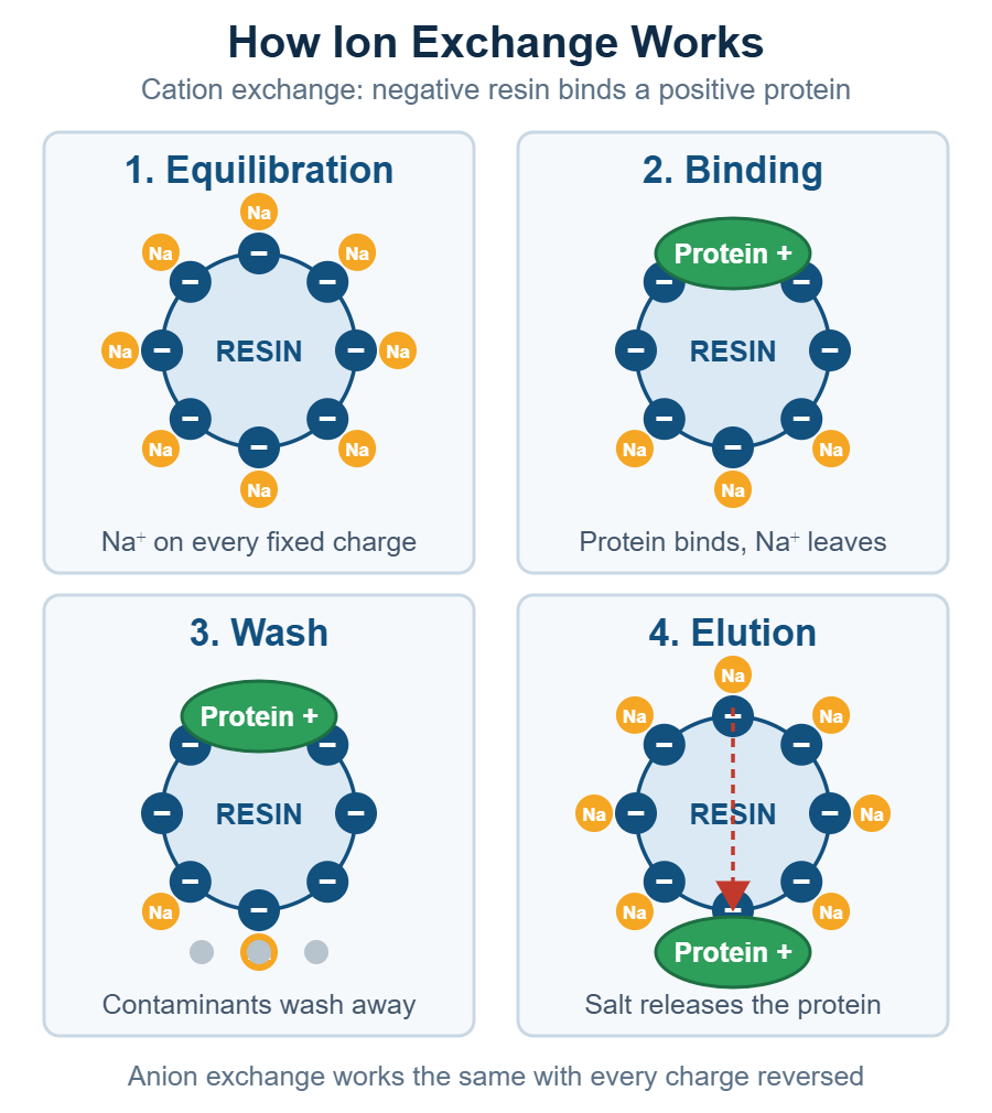
IEX works in four stages: equilibration, binding, washing, and elution, shown in Figure 1. The resin’s fixed charges start paired with small counterions. When you load your sample, analytes with a stronger charge interaction displace those counterions and take their place. Species without a sufficiently favorable charge interaction generally pass through, while oppositely charged species that interact strongly enough are retained. You then break the interaction and collect what comes off.
Equilibration. You flush the column with a low-ionic-strength start buffer at your chosen pH. This sets the resin’s charge state and loads it with exchangeable counterions — typically Na⁺ for a cation exchanger, Cl⁻ for an anion exchanger.
Binding. The sample goes on in that same low-salt buffer. Analytes compete with the counterions for the fixed charges. Competition strength depends on charge density and how closely the molecule can approach the surface, which is why two proteins with the same net charge can still separate.
Washing. Still at low salt, you flush out anything that did not bind. This is where most of your contaminant removal happens.
Elution. You raise the ionic strength — usually a linear NaCl gradient — so the incoming salt ions outcompete the bound analytes. Weakly bound species let go first, strongly bound species last. The alternative is a pH gradient that neutralizes the analyte’s charge instead of competing it off.
Anion vs cation exchange: what’s the difference?
The difference is which charge the resin carries, and therefore which charge it captures, as summarized in Table 2. Anion exchangers carry positive fixed groups and bind negatively charged analytes. Cation exchangers carry negative fixed groups and bind positively charged analytes. The naming trips people up constantly: the exchanger is named for the ion it binds, not the charge it carries.
| Cation exchange (CEX) | Anion exchange (AEX) | |
|---|---|---|
| Fixed charge on resin | Negative | Positive |
| Binds | Cations (+) | Anions (−) |
| Typical target | Basic proteins, amino-rich surfaces | Acidic proteins, carboxyl-rich surfaces |
| Strong exchanger group | Sulfonate / sulfopropyl (S, SP) | Quaternary ammonium (Q) |
| Weak exchanger group | Carboxymethyl (CM) | Diethylaminoethyl (DEAE) |
| Working buffer pH | Below the analyte’s pI | Above the analyte’s pI |
| Common counterion | Na⁺ | Cl⁻ |
The mnemonic that actually sticks: a cation exchanger is a cation catcher. It must be negative to catch a positive.
Which one do you need?
Your protein’s pI is the starting point. Stability window, contaminant charge and sample conductivity all narrow the choice from there. A protein carries a net positive charge below its pI and a net negative charge above it. So:
- Working below the pI → protein is positive → use a cation exchanger
- Working above the pI → protein is negative → use an anion exchanger
A protein with a pI of 5.5 is negatively charged at pH 8 and will bind an anion exchanger. The same protein at pH 4 is positively charged and binds a cation exchanger. Both are valid routes to the same purification — the choice usually comes down to which pH your protein tolerates and where your contaminants sit.
Strong vs weak ion exchangers — and when to use each
“Strong” and “weak” describe how the exchanger’s charge responds to pH — not how tightly it binds. A strong exchanger stays fully charged across essentially the whole working pH range. Table 3 gives the groups and their charged ranges. A weak exchanger progressively loses charge as pH approaches the pKa of its functional group, and with it, loses capacity.
| Exchanger | Charged range | Type | Group |
|---|---|---|---|
| Q | Fully charged, pH 2–122 | Strong anion | Quaternary ammonium |
| DEAE | Charged below ~pH 91,2 | Weak anion | Diethylaminoethyl |
| SP / S | Fully charged, pH 4–132 | Strong cation | Sulfopropyl / sulfonate |
| CM | Charged above ~pH 61,2 | Weak cation | Carboxymethyl |
The carboxylate group on a CM exchanger has a free-ligand pKa near 4–5, but that is not the number to design around. Manufacturers specify CM resins for use above about pH 6, because charge on the bead does not develop fully until well above that value.2 Cytiva gives a working range of pH 6–10 for CM.1
Use a strong exchanger when you need predictable, pH-independent capacity, you are working near the extremes of the pH range, or you are developing a method and want one fewer variable. Q and SP are reasonable starting points when screening a new method.
Use a weak exchanger when you want extra selectivity, or you need gentler elution conditions. Because a weak exchanger’s charge varies with pH, you can elute by shifting pH rather than piling on salt — useful for proteins that aggregate or precipitate at high ionic strength.
The practical failure mode: running CM at pH 5.0 and wondering why capacity is so low. Nothing has failed — at pH 5.0 the carboxylates are only partly ionized, so that capacity was never there.
How to choose your exchanger and buffer pH
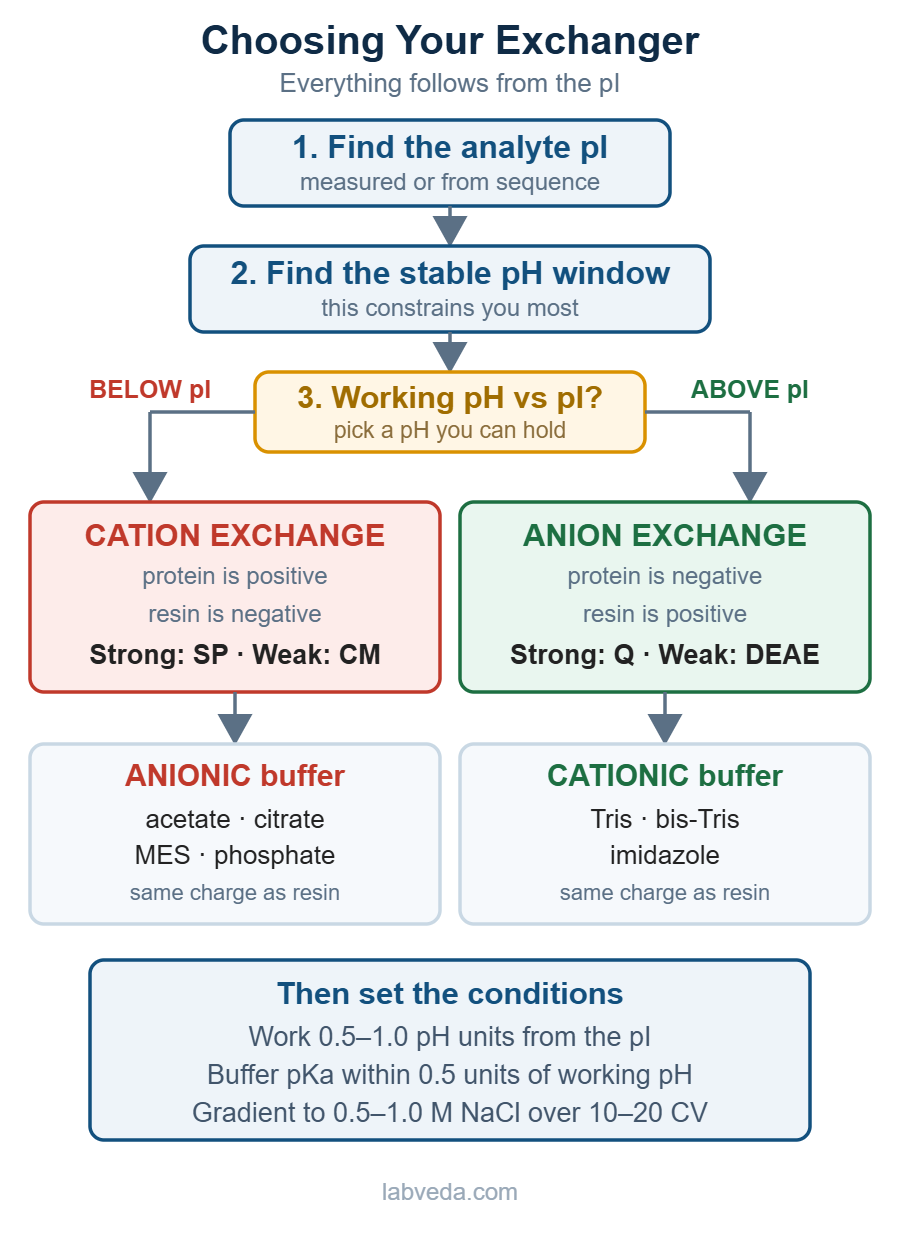
The whole decision follows from the pI. Figure 2 maps it; the seven steps below work through it in order.
Work through these seven steps in order.
-
Find your analyte’s pI
Measure it, or calculate it from sequence. Everything downstream depends on this number.
-
Establish your protein’s stable pH window
There is no point picking an ideal binding pH if your protein denatures there. This constraint usually narrows the options more than anything else.
-
Pick the exchange mode
Choose a working pH inside the stable window, then apply the rule: pH above pI → anion exchanger; pH below pI → cation exchanger. If both are viable, prefer the one where your main contaminants carry the opposite charge, so they wash through instead of co-binding.
-
Set the pH offset from pI
Work at least 0.5–1.0 pH unit away from the pI. Sitting at the pI means near-zero net charge and no binding. Further from the pI means stronger binding — good for capture, potentially too tight for a clean elution.
-
Choose a strong exchanger unless you have a reason not to
Q for anion, SP for cation. Verify your working pH sits inside the resin’s charged range.
-
Match the buffering ion to the resin
The buffering ion should carry the same charge as the resin’s fixed groups, so it does not take part in the exchange. Cation exchange (negative fixed groups) → use an anionic buffer: acetate, citrate, MES, phosphate, PIPES. Anion exchange (positive fixed groups) → use a cationic buffer: Tris, bis-Tris, imidazole, triethanolamine, piperazine. Buffering capacity is greatest close to the pKa and remains useful to roughly ±1 pH unit; for IEX, vendors recommend staying within about 0.5–0.6 units of the working pH.
-
Set a screening gradient
As a starting condition, run a linear gradient from low ionic strength to 0.5–1.0 M NaCl over 10–20 column volumes. Adjust for your resin, feed conductivity, binding strength and the resolution you need — this is a screening point, not a fixed recipe.
A worked example: a protein with pI 5.5
Say you have a 45 kDa protein, pI 5.5, stable between pH 5 and pH 9, and your main contaminant is a basic protein with pI around 8.5.
Mode. Both routes are technically open, but only one is sensible. Running cation exchange means working below pH 5.5, and your protein is only stable down to pH 5 — a 0.5 unit window with no margin. Anion exchange means working above pH 5.5, and you have room all the way to pH 9. Choose anion exchange.
Contaminant. At pH 7.5 your target (pI 5.5) is comfortably negative and binds. The contaminant (pI 8.5) is net positive at pH 7.5, so it should not bind an anion exchanger and is expected in the flow-through. One buffer choice removes your main impurity before the gradient even starts. Confirm it experimentally rather than assuming it: a net-positive protein can still bind an anion exchanger through a localized patch of negative surface charge, which is why some antibodies bind both exchangers at the same pH.
Resin. Q for a first pass. It is fully charged at pH 7.5, so capacity will not drift if your buffer prep varies slightly. DEAE would also be charged here, but its capacity falls off as you approach pH 9 and there is no reason to accept that variable on a first method.
Buffer. Anion exchange needs a cationic buffer. Tris is the obvious candidate, with a pKa of 8.06 at 25 °C.4 Be honest about the margin though: buffering capacity is strongest near the pKa and is generally useful to about ±1 pH unit, which puts pH 7.5 at the weak end of the useful range for Tris.8 It works at 20 mM Tris-HCl, but buffering capacity is thin. Running at pH 8.0 gives you more room, and bis-Tris propane (pKa 6.8 and 9.0) or triethanolamine (pKa ≈ 7.8) is a better match if you specifically need pH 7.5.4 If you are titrating buffers by hand, our 37% HCl molarity guide and 50% NaOH molarity guide cover the stock calculations. Phosphate breaks the rule. The same-charge rule argues against it on an anion exchanger, yet phosphate is used with anion exchange routinely and vendors name it as an explicit exception. If you do use it, prepare it carefully — batch-to-batch variation in phosphate buffers is a real reproducibility risk.
Temperature. This matters more than people expect. Tris has one of the largest temperature coefficients of any common buffer, around −0.03 pH units per °C.5,6 A Tris buffer titrated to pH 7.5 on the bench at 22 °C sits near pH 8.0 in a 4 °C cold room — a bigger shift than the margin this whole example depends on. Titrate your buffers at the temperature you will actually run them.
Gradient. Equilibrate in 10 column volumes of buffer, or until UV, pH and conductivity baselines are stable. Load, wash until the baseline returns, then run a linear gradient to 0.5 M NaCl over 20 column volumes, followed by a 1 M strip.
If nothing binds. Check the sample’s conductivity and pH against the start buffer before anything else. Conductivity is the more common cause, especially when the load has come straight off a salt elution or a concentrated stock — a sample carried over from a previous high-salt step will flow straight through a correctly set-up column. pH is the usual culprit when the sample was never buffer-exchanged.
What elutes first in ion exchange chromatography?
The most weakly bound species elutes first. In a salt gradient, molecules release in ascending order of interaction strength — weakest charge interaction off the column first, strongest last. Anything that did not bind at all comes off in the flow-through, before the gradient even starts.
Interaction strength is not simply net charge. It is governed by charge density and accessibility — how much charge is concentrated on the surface that actually contacts the resin. A protein with a modest net charge but a strong, exposed charge patch can outbind a protein with a higher net charge spread thinly over a large surface. This is why IEX resolves proteins that look identical by net charge alone, and why elution order sometimes defies the simple prediction.
In a pH gradient the logic differs: species elute as the pH passes through the point where their net charge neutralizes, so elution order broadly tracks pI. Two caveats worth knowing — elution typically begins around 0.5 pH units before the pI is reached, and the correlation degrades for proteins with a pI between 6 and 8, which tend to elute well above their pI. If pI-based separation is the goal, chromatofocusing is more reliable than an IEX pH gradient, and watch for precipitation as proteins pass through their pI.
Binding capacity and scaling up
IEX scales more predictably than most modes, but only if you size the column on the right number. The figure that matters is dynamic binding capacity (DBC) under your conditions. Datasheets quote either static binding capacity, measured with excess protein and no flow, or a DBC measured with a model protein such as BSA at a fixed residence time — neither is your feed. Total ionic capacity, quoted in mmol/mL, is a different number again: it counts charged groups, not bound protein.
DBC falls as flow rate rises, because large molecules need time to diffuse into the pores.3 It also falls as you move the working pH closer to the analyte pI, since net charge drops. A resin quoted at 50 mg/mL may deliver half that under your conditions. How dynamic binding capacity trades off against residence time, and what pore size, particle size and ligand density each cost you across resin types, is worked through in the chromatography resins guide.
Two habits keep scale-up honest:
- Measure DBC yourself at 10% breakthrough, under the flow rate and buffer you intend to run.3,7 Check the breakthrough criterion before comparing datasheets — some vendors quote 50% breakthrough, which makes a resin look far better than one measured at 10%.
- Scale by column volume and bed height, not diameter alone. Keep bed height and linear flow rate constant and increase diameter — that preserves residence time, which is what governs binding.
A common industry practice is to load to about 80% of the DBC measured at 10% breakthrough, leaving a safety margin for variation between resin lots, cycles and batches.7 That figure protects against breakthrough, not resolution. Where separation matters more than throughput, the guidance from Cytiva for gradient elution is far more conservative: no more than about 30% of total binding capacity — a different denominator from the DBC figure above, so the two are not directly comparable.1 Load closer to capacity and you buy throughput at the price of yield the first time anything drifts.
Common IEX problems and how to fix them
Table 4 lists the eight symptoms that account for most bad IEX runs, with the likely cause and fix for each.
| Symptom | Likely cause | Fix |
|---|---|---|
| Nothing binds — everything in flow-through | Working pH on the wrong side of the pI, or too close to it | Recheck pI. Move working pH 0.5–1.0 units further from it, or switch exchange mode |
| Nothing binds, pH is correct | Sample ionic strength too high | Desalt or dilute the sample into start buffer before loading. Buffer exchange or a size exclusion step both work |
| Weak binding, low capacity | Weak exchanger operating outside its charged range | Check resin pKa vs working pH. CM below pH 6 or DEAE above pH 9 will underperform. Switch to SP or Q |
| Peak tailing | Overloading, or secondary hydrophobic interactions | Reduce load. Confirm the buffer pKa is close to the working pH. See our diagnostic path for tailing peaks |
| Broad, smeared peaks | Gradient too shallow, or column poorly equilibrated | Steepen gradient. Extend equilibration to 10+ column volumes |
| Co-elution / poor resolution | Gradient too steep | Shallow the gradient across the region of interest |
| Capacity falling run over run | Incomplete regeneration, fouling | Run a high-salt strip and CIP between cycles. See column cleaning |
| Retention drifting between runs | Buffer pH drift or inconsistent preparation | Prepare buffers fresh, verify pH at working temperature. See drifting retention times |
Buffer pH is the first thing to check for the binding failures. If you are also chasing peak shape problems more generally, our guide to HPLC peak shape troubleshooting covers the causes that are not charge-related.
Applications of ion exchange chromatography
IEX turns up wherever charge is a usable handle:
- Protein and enzyme purification — the dominant use, from lab-scale prep to manufacturing, often paired with a size exclusion polishing step
- Monoclonal antibody polishing — charge-variant separation and aggregate removal
- Nucleic acid purification — the phosphate backbone gives DNA and RNA a strong, consistent negative charge, making anion exchange a natural fit
- Water treatment and deionization — the large-scale industrial application
- Food and beverage — sugar refining, organic acid removal
- Clinical analysis — HbA1c determination is a routine cation exchange assay
Frequently asked questions
What is ion exchange in chromatography?
Ion exchange is the reversible swapping of ions between the mobile phase and charged groups fixed to the stationary phase. Analytes displace small counterions to bind, then get displaced in turn when salt concentration rises.
What is the purpose of ion exchange chromatography?
To separate or purify molecules by net charge. It offers high binding capacity and pH-tunable selectivity, which makes it a common capture or polishing step. For monoclonal antibodies, Protein A affinity is usually the capture step and IEX is used for polishing.
What elutes first in ion-exchange chromatography?
The most weakly bound species. Unbound material appears in the flow-through, then bound species elute in ascending order of interaction strength as salt concentration increases.
What is the principle of ion exchange chromatography?
Molecules carrying a charge opposite to the resin’s fixed groups bind electrostatically. Binding strength depends on charge density and accessibility. Raising ionic strength or shifting pH reverses the interaction and releases them in order.
What are the different types of ion exchange chromatography?
Two, by charge: anion exchange (positive resin, binds anions) and cation exchange (negative resin, binds cations). Each subdivides into strong exchangers (Q, SP) and weak exchangers (DEAE, CM).
What is the difference between anion and cation exchange?
An anion exchanger carries positive fixed groups and binds negatively charged analytes, working above the analyte’s pI. A cation exchanger carries negative fixed groups and binds positively charged analytes, working below the pI.
What are the advantages of ion exchange chromatography?
High loading capacity, good scalability, mild non-denaturing conditions, pH-based selectivity control, and resins that regenerate for many cycles.
What are the limitations of ion exchange chromatography?
Samples must be low in ionic strength before loading, which often means an extra desalting step. It only separates molecules that differ in charge, and high salt eluates frequently need buffer exchange downstream.
What is the difference between ion exchange chromatography and ion chromatography?
Ion exchange chromatography is the general separation mode. Ion chromatography (IC) is a specific analytical application of it, typically small inorganic ions with conductivity detection.
Is IEX the same as IEC?
Yes. IEX is the more common abbreviation in industry and bioprocessing; IEC is more common in academic and textbook usage. Both mean ion exchange chromatography.
References
- Cytiva — Ion Exchange Chromatography: Principles and Methods (handbook)
- Cytiva — HiTrap DEAE FF, CM FF, Q FF, SP FF and ANX FF — Instructions for Use (71501751 AK, 2020)
- Cytiva — Capto S, Capto Q and Capto DEAE — data file CY13280-24Aug20-DF (2020)
- Merck / Sigma-Aldrich — Buffer Reference Center — useful pH ranges of selected biological buffers (25 °C, 0.1 M)
- MilliporeSigma — Biological Buffers Selection Guide (MS_PS3138EN Ver. 2.0, 2019)
- Promega — Temperature Dependence of pH for Commonly Used Buffers (Technical Reference GE647, 2010)
- Challener, C. A. — Determining and Optimizing Dynamic Binding Capacity. BioPharm International 2023;36(4):10–14
- Chemistry LibreTexts — Practical Aspects of Buffers — buffer range (pKa ± 1)
Further reading
- Phenomenex — Principles of Ion Exchange
- Cytiva — Troubleshooting Protein Loss During IEX Chromatography
- Merck / Sigma-Aldrich — Practical Considerations for IEX Separation
- Thermo Fisher Scientific — Tech Tip #62: Ion Exchange Chromatography